
The third day of CICON19 opened with session 7 devoted to tumor antigens, with a talk by Yardena Samuels, PhD, of the Weizmann Institute of Science (Israel), who discussed her work covering a variety of issues relating to tumor antigens. Samuels shared how tumor heterogeneity—where protein expression differs throughout the tumor—affects immune responses against cancer. After noting that tumor mutational burden (TMB) is not a sufficient predictor of anti-tumor immune responses, she discussed how subjecting a melanoma cell line to type B ultraviolet radiation increased TMB as well as tumor heterogeneity and made these tumors less responsive to anti-PD-1 immunotherapy. However, when she took single clones from these UV-irradiated tumors and grew them into tumors, they had reduced heterogeneity and were more sensitive to T cell infiltration and immune-mediated elimination. She determined that tumor diversity is an important factor in clinical responses to immunotherapy, and suggested that minimizing tumor heterogeneity might serve to improve immune detection of reactive tumor neo-antigens.
Yardena Samuels, PhD, discusses tumor antigens.
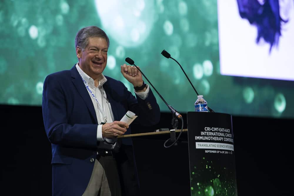
Next, Robert D. Schreiber, PhD, an associate director of the Cancer Research Institute (CRI) Scientific Advisory Council at the Washington University School of Medicine, discussed his recent discoveries regarding the importance of class II MHC-restricted (MHC-II) mutated neoantigens in enabling the immune system to eliminate tumors with the help of immunotherapy. Specifically, these MHC-II neoantigens were able to promote the priming of “killer” T cells (which bind MHC-I neoantigens) and stimulate their cancer-killing activity. Schreiber showed that vaccines incorporating both MHC-I and MHC-II neoantigens were more effective than those that with only MHC-I neoantigens. In this regard, one type of each neoantigen—so long as they were expressed homogeneously in tumors—was sufficient to render these tumors susceptible to immunotherapy. Lastly, Schreiber demonstrated that even when tumors only expressed MHC-I neoantigens, the activity of the “helper” T cells (which bind MHC-II neoantigens) were required for immunotherapy’s effectiveness, most likely through their ability to stimulate “killer” T cells to target tumors via MHC-I neoantigens.
Robert D. Schreiber, PhD, discusses MHC-II-restricted mutated neoantigens.
Following Schreiber was Chloe Chong, a PhD student at the Ludwig Cancer Research Centre at the University of Lausanne, who focused on efforts to expand the repertoire of tumor markers that could be targeted through immunotherapy. In addition to the already recognized categories of tumor-associated antigens (which include cancer-testis, overexpressed normal, and differentiation antigens) and tumor-specific antigens (which include oncoviral as well as shared and private neoantigens), Chong sought to identify potential targets that arise from gene fusions, RNA editing, transcript-alternative splicing, and modifications that are made to proteins after they’re produced, among other processes. To do so, her team has been using a new computational model called NewAnce (New Approach for Non-Canonical Element identification) that combines two mass spectrometry tools—MaxQuant and Comet—in order to identify targetable antigens associated with the protein-coding regions as well as the non-canonical regions of the genome.
Chloe Chong discusses expanding the repertoire of tumor markers.
Next, former CRI grantee Sjoerd van der Burg, PhD, of Leiden University (The Netherlands), who presented his findings relating to why some patients with human papilloma virus (HPV)-positive oral cancer relapse after initially responding to an HPV-targeting vaccine in combination with PD-1 immunotherapy. In subsequent experiments with mice, van der Burg found that this relapse was associated with suboptimal T cell responses, although neither PD-1 immunotherapy nor agonistic treatments targeting the OX40 and 4-1BB pathways could protect against relapse. He also found that these relapsed tumors became “cold” and were characterized by increased TGF-b activity. Promisingly, when cisplatin chemotherapy was administered after initial tumor regression, it sensitized these tumors to subsequent vaccination, resulting in superior tumor control that was associated with the restoration of myeloid immune cell infiltration.
Sjoerd van der Burg, PhD, discusses HPV-positive oral cancer relapse.
In addition to chemotherapy, radiation therapy is another promising option to use in combination with immunotherapy, as it can kill cancer cells and cause them to release their antigens, creating a vaccine-like effect. Not only can this alert the immune system to the cells in the irradiated tumor, but it can also stimulate the immune system to seek out and eliminate tumor cells elsewhere in the body, a phenomenon known as the abscopal effect. However, as Lorenzo Galluzzi, PhD, of Weill Cornell Medical College, showed, if cancer cells die via autophagy—a type of cellular recycling process—this can inhibit abscopal responses. In particular, he found that this occurs as a result of the tidy disposal of mitochondria within cells. This, in turn, limits the potential activity of the cGAS/STING signaling pathway that would otherwise be stimulated by the presence of mitochondrial DNA and the production of type I interferon molecules.
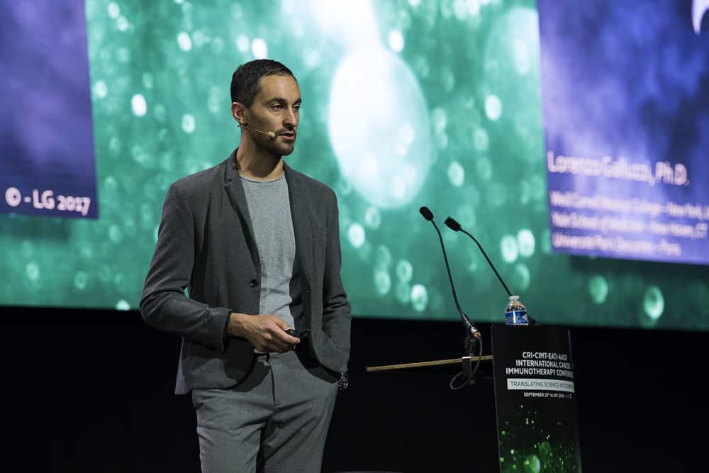
Lorenzo Galluzzi, PhD, discusses inhibiting abscopal responses.
Session 8: Vaccination Strategies
Sandra Demaria, MD, of Weill Cornell Medical College, also spoke about these “on site” vaccine-like capabilities of radiation therapy. In lung cancer patients treated with the combination of radiation therapy and CTLA-4 checkpoint immunotherapy, clinical responses were associated with increased levels of interferon-b as well as significant changes in the T cell receptor (TCR) repertoire and, in at least one complete responder, the expansion of tumor-derived clones in the blood. In subsequent preclinical experiments, Demaria showed that this increased diversity and expansion of the TCR repertoire was driven primarily by the radiation treatment.
Sandra Demaria, MD, discusses vaccine-like capabilities of radiation therapy.
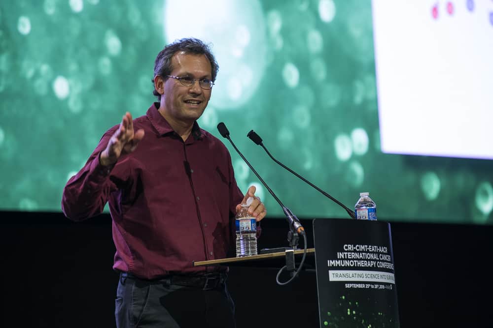
The next two speakers—Sebastian Kreiter, MD, and Özlem Türeci, MD, both of BioNTech (Germany)—spoke about their company’s efforts involving RNA-based cancer vaccines. Kreiter began by explaining how these RNA vaccines can either target dendritic cells locally (via ultrasound-guided injection into lymph nodes) or systemically (via intravenous injection of liposomal complexes within which the RNA vaccines have been incorporated).
Sebastian Kreiter, MD, discusses RNA-based cancer vaccines.
Preclinically, Kreiter revealed the synergistic benefits of this latter approach when it was combined with local tumor radiation in preclinical models, whereas Türeci revealed promising early clinical results. In a trial involving patients with advanced melanoma, an RNA lipoplex vaccine designed to target four tumor-associated antigens—tyrosinase, MAGE-A3, NY-ESO-1, and TPTE—produced new responses or enhanced existing T cell responses against at least one of these targets in almost all patients, and demonstrated clinical activity in several patients. The vaccine was especially effective at stimulating immune responses against the NY-ESO-1 and MAGE-A3 targets, which were observed in 80% of patients. Additionally, in patients who previously failed to respond to PD-1 checkpoint immunotherapy, there was evidence that this vaccine may be able to re-sensitize patients to this approach when used in combination.
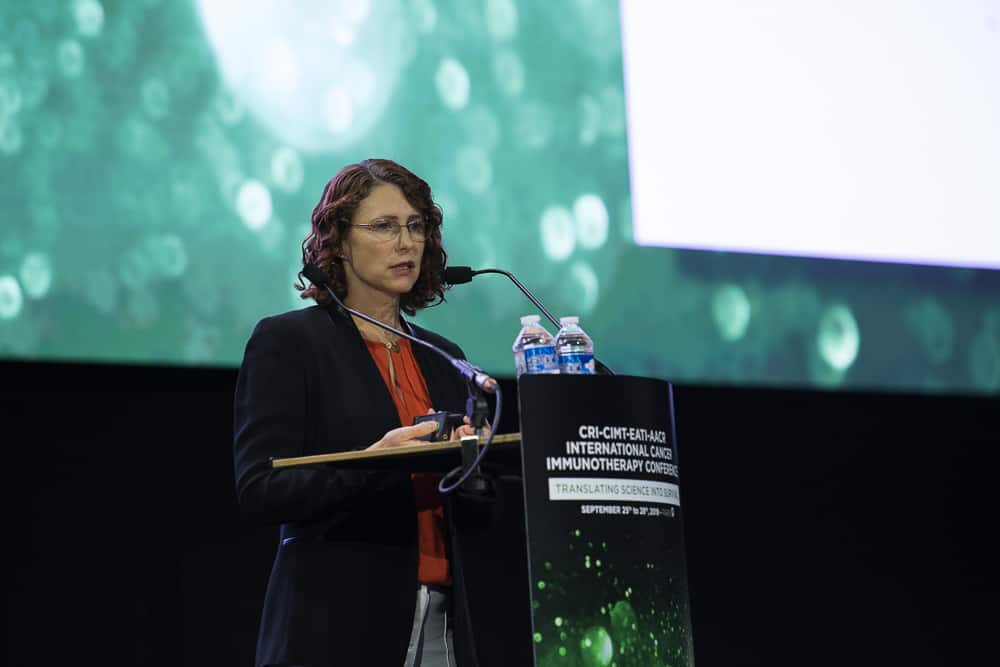
Özlem Türeci, MD, discusses RNA-based cancer vaccines.
Elizabeth Evans, PhD, of Vaccinex, next discussed combination immunotherapy involving a SEMA4-blocking antibody, which is designed to reverse myeloid cell-mediated immunosuppression and promote the infiltration and activity of tumor-targeting T cells. Preclinically, the addition of the anti-SEMA4D antibody improved the effectiveness of immunotherapies targeting the PD-1, CTLA-4, LAG-3, and TGF-b pathways. More recently, this approach was applied clinically in combination with PD-L1 immunotherapy in patients with advanced lung cancer who were previously treated with, but did not respond to, PD-1 or PD-L1 checkpoint immunotherapy. Interim results from this trial showed that more than half of the seventeen patients experienced disease control, while two had partial responses.
Elizabeth Evans, PhD, discusses combination immunotherapy involving a SEMA4-blocking antibody.
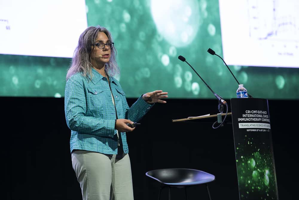
Next, Michelle R. Brault, PhD, of the Fred Hutchinson Cancer Research Center, spoke about a novel target—an antigen known as PBK—that could potentially be used to treat pediatric brain cancer. Normally expressed in testis and fetal tissues, this protein is upregulated in several types of cancers, including childhood brain cancer. After isolating PBK-targeting “killer” T cells, Brault found that they can recognize and kill pediatric brain cancer cells in the lab, suggesting that targeting PBK in the clinic might be a valuable option, especially with adoptive cell immunotherapy approaches.
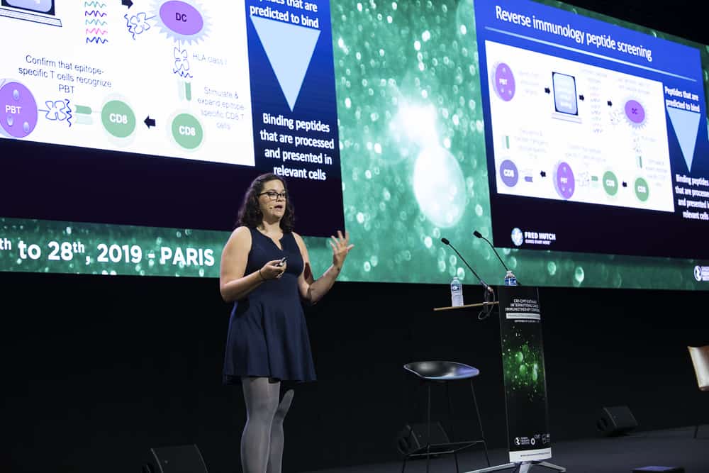
Michelle R. Brault, PhD, discusses PBK's potential in pediatric brain cancer.
Most current strategies look for potential targets for immunotherapy in the protein-coding regions of our genome. However, according to Tamara Ouspenskaia, PhD, of the Broad Institute of MIT and Harvard, regions of the genome known as “unannotated open reading frames” that haven’t previously been catalogued might be an untapped source of targets for cancer immunotherapy. In work involving CRI postdoctoral fellow Susan Klaeger, PhD, Ouspenskaia showed that ribosome profiling was able to identify which of these open reading frames might be expressed, regardless of whether they had been previously catalogued. Upon deeper examination, their team identified thousands of potential targets that were derived from these regions and displayed by cancer cells via the MHC system. Moving forward, their goal is to improve our understanding of the impact of these proteins in cancer and to see whether targeting them through immunotherapy might have clinical value.
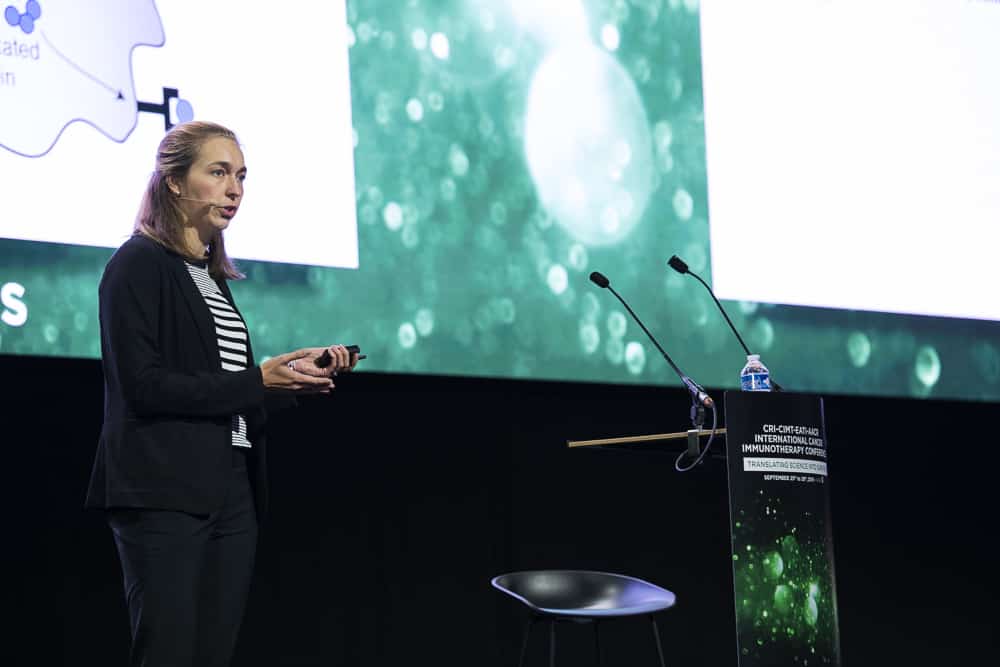
Tamara Ouspenskaia, PhD, discusses unannotated open reading frames.
Session 9: New Trends in Technology and Informatics
The first speaker of the next session was Hannah Carter, PhD, of the University of California, San Diego, who discussed how the specific versions of the MHC molecules a person possess determines how their cancer’s mutations are presented to the immune system. Interestingly, mutations that aren’t efficiently presented by a particular person’s MHC molecules are more likely to be observed and persist because their immune system won’t have a chance to recognize it and launch a response against the cancer cells that express it. Some mutations, Carter showed, are commonly seen across a variety of tumor types due to the fact that they are poorly presented in the majority of patients. With respect to mutations that drive cancerous behavior, if they are efficiently presented, the potentially cancerous cells that express them can be better recognized and eliminated by the immune system. In addition to possibly delaying the onset of tumor development, the presence of these efficiently presented driver mutations also makes patients more likely to respond to checkpoint immunotherapy.
Hannah Carter, PhD, discusses MHC molecules.
Next, E. John Wherry, PhD—a former CRI postdoctoral fellow who is now a member of CRI’s Clinical Accelerator leadership—of the University of Pennsylvania, spoke about the molecular origins of exhausted “killer” T cells. In work involving CRI postdoctoral fellow Josephine R. Giles, PhD, Wherry discussed the epigenetic landscape of T cells in order to understand what influences the durability of responses in patients treated with immunotherapy. What they found is that TOX, a gene transcription factor, serves as a primary driver of the development of exhausted T cells by epigenetically programming T cells in both chronic infection and cancer. In addition to being required for the development of these exhausted T cells, TOX also prevents T cells from becoming terminally differentiated (fully mature) effector T cells. Therefore, Wherry proposed that these TOX-induced epigenetic changes might serve as valuable targets that may have therapeutic potential to improve the effectiveness and durability of immunotherapy.
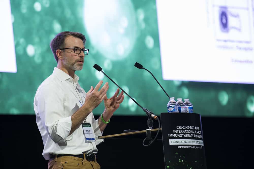
E. John Wherry, PhD, discusses the molecular origins of exhausted "killer" T cells.
As with earlier talks by Chong and Carter, Lausanne University Hospital’s Michal Bassani-Sternberg, PhD, also focused on identifying targetable antigens that lie outside of the canonical protein-coding regions of our genome. To search this space effectively, she stressed the necessity of advanced analytical pipelines to control for potential erroneous results due to the relative rarity of these molecular targets—only four were seen to be presented via the class II MHC system. Moving forward, she plans to focus on determining whether or not these targets might be relevant for immunotherapy, how their production is regulated, and whether tumors might be able to escape immune responses that target these molecules under selective pressure.
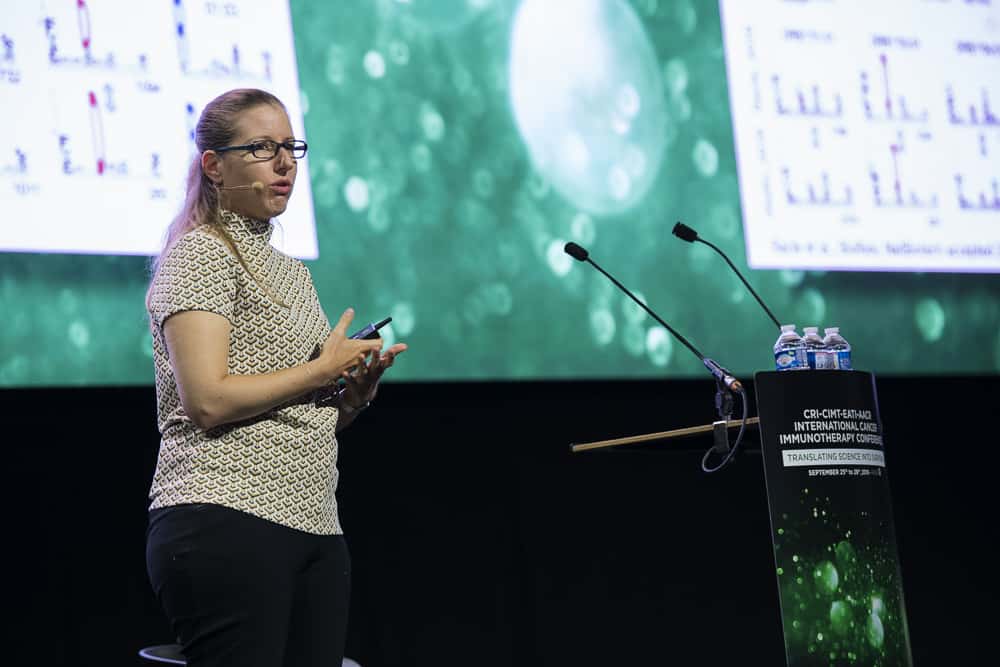
Michal Bassani-Sternberg, PhD, discusses molecular targets outside of the canonical protein-coding regions of our genome.
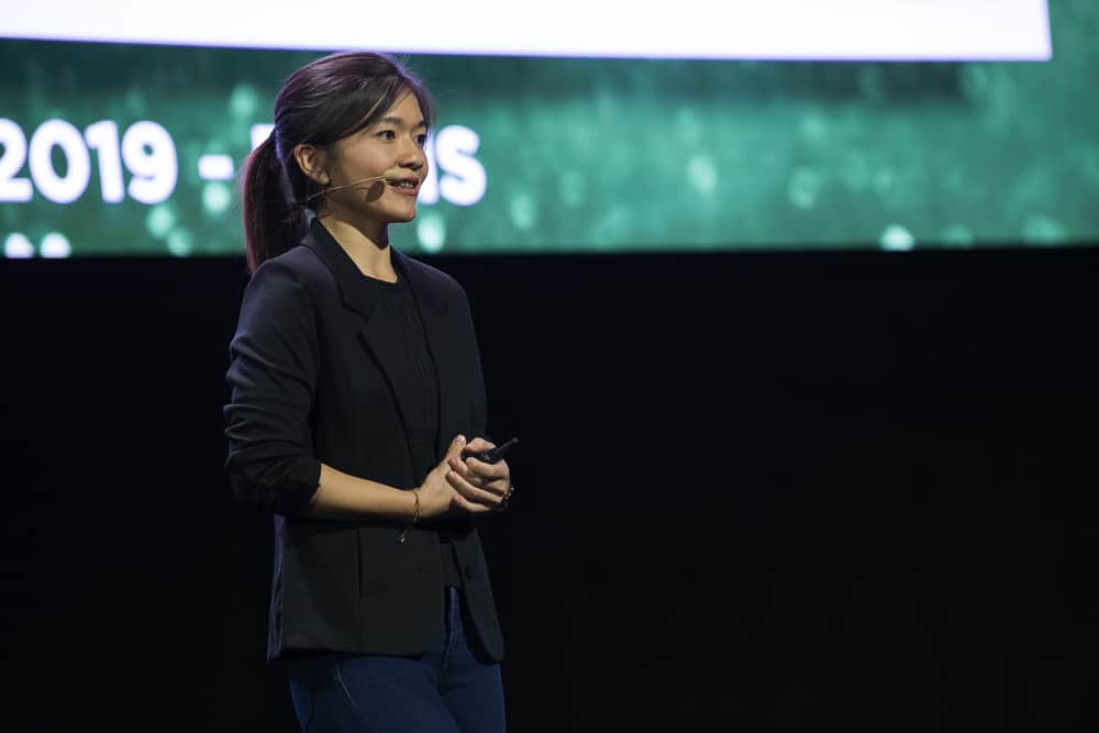
The following speaker—CRI postdoctoral fellow Livnat Jerby, PhD, of the Broad Institute—discussed the forces that shape cancer-promoting programs in synovial sarcoma. While sarcomas typically express antigens capable of stimulating the immune system, they are often “cold” tumors from which immune cells are excluded and are therefore typically unresponsive to immunotherapy. Jerby found that the SS18-SSX oncogenic fusion protein directly regulates this core cancer-promoting program, which is associated with poor clinical outcomes. However, by inhibiting both HDAC and CDK4/6—two of the mediators of this SS18-SSX-driven program—Jerby showed that its ability to promote cancer development could be blocked, and Jerby and her team are now exploring whether or not this approach might sensitize these sarcoma tumors to checkpoint immunotherapy.

Livnat Jerby, PhD, discusses the forces that shape cancer-promoting programs in synovial sarcoma.
Session 10: Tumor Microenvironment Analysis
Wolf Hervé Fridman, MD, PhD, of the Centre de Recherche des Cordeliers (France), started off the next and final session of the third day of CICON19 with a discussion of the important role that B cells play within tumors and how their presence is linked to cancer patient outcomes. Fridman began with soft tissue sarcomas, which he grouped into categories based on their immune characteristics. In the most prominent “immune-high” group, half the patients responded to PD-1 immunotherapy, compared to none in the two “immune-cold” groups (which were the most common and comprised roughly half of all cases). Fridman found that the immune-high group was defined by the presence of important immune related-structures known as tertiary lymphoid structures, which could be used as a surrogate indicator to predict patient responses to immunotherapy. Interestingly, B cell signatures were the best predictor of overall survival and were the dominant signature regardless of whether or not tumors had high or low levels of “killer” T cells.
Wolf Hervé Fridman, MD, PhD, discusses the role that B cells play within tumors.
In kidney cancer, however, Fridman found that B cells could aid the progression of cancer. In these tumors, high levels of the complement component C1q caused B cells to produce antibodies that helped activate the classical complement pathway. Normally, complement helps the immune system to function, but in this context is was associated with poor clinical outcomes. Due to the expression of certain complement inhibitors, cancer cells themselves were protected from complement-mediated destruction, and instead the complement activation resulted in tumor-enhancing inflammation and blood vessel growth.
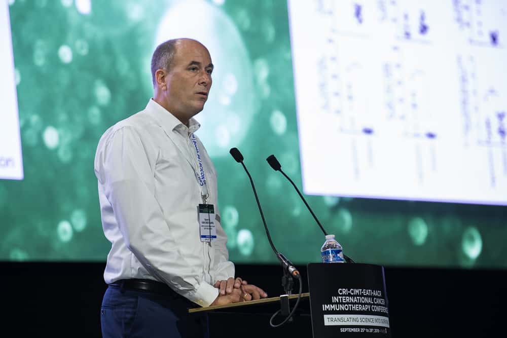
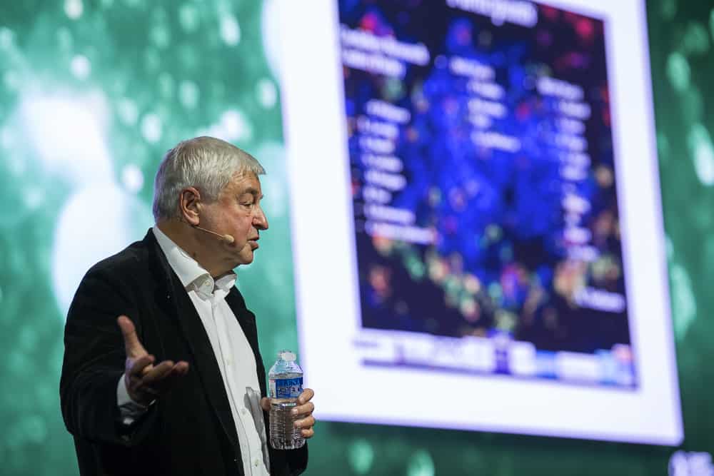
Next, CRI CLIP Investigator Nir Hacohen, PhD, of Massachusetts General Hospital and the Broad Institute, discussed his efforts to improve our ability to predict potential neoantigen targets expressed by cancer cells. By sequencing single alleles of HLA (an important component of the MHC system that binds and presents antigens), Hacohen was able to determine that similarities in antigen motifs corresponded with similarities in the sequences of these different HLA proteins in the context of the class I MHC molecules.
Nir Hacohen, PhD, discusses the prediction of potential neoantigen targets.
Additionally, he was able to characterize the various lengths of the antigen fragments that are bound by various HLAs, and observed that the rules of protein cleavage—the process by which proteins are “chopped up” into smaller bits to be presented via HLA/MHC—were preserved across various cell types, including cancer cells. Surprisingly, even when cancer cells were stimulated by interferon gamma, this signature remained unchanged.
Lastly, Hacohen showed that incorporating cell-intrinsic processes such as protein cleavage and gene expression significantly improved the predictive power of his mass spectrometry-based integrative models, which were able to outperform today’s state-of-the-art predictors with respect to identifying antigens presented by tumors cells. Fortunately, Hacohen announced, these tools for visualization, exploration, and prediction will all be freely available after their forthcoming paper is published.
The next speaker—Miriam Merad, MD, PhD, of the Icahn School of Medicine at Mount Sinai and a winner of the 2018 CRI William B. Coley Award—discussed the theory that there are two distinct lineages of macrophages. The first are tissue-resident macrophages that self-renew in tissues and are imprinted by tissue-specific cues. The second are bone marrow-derived macrophages that are recruited from the blood in response to injury or inflammation. Although macrophages are the most abundant immune cells within tumors, targeting them has proven difficult, Merad suggested, because our knowledge is mostly based on bone marrow-derived macrophages that are generated outside the body.
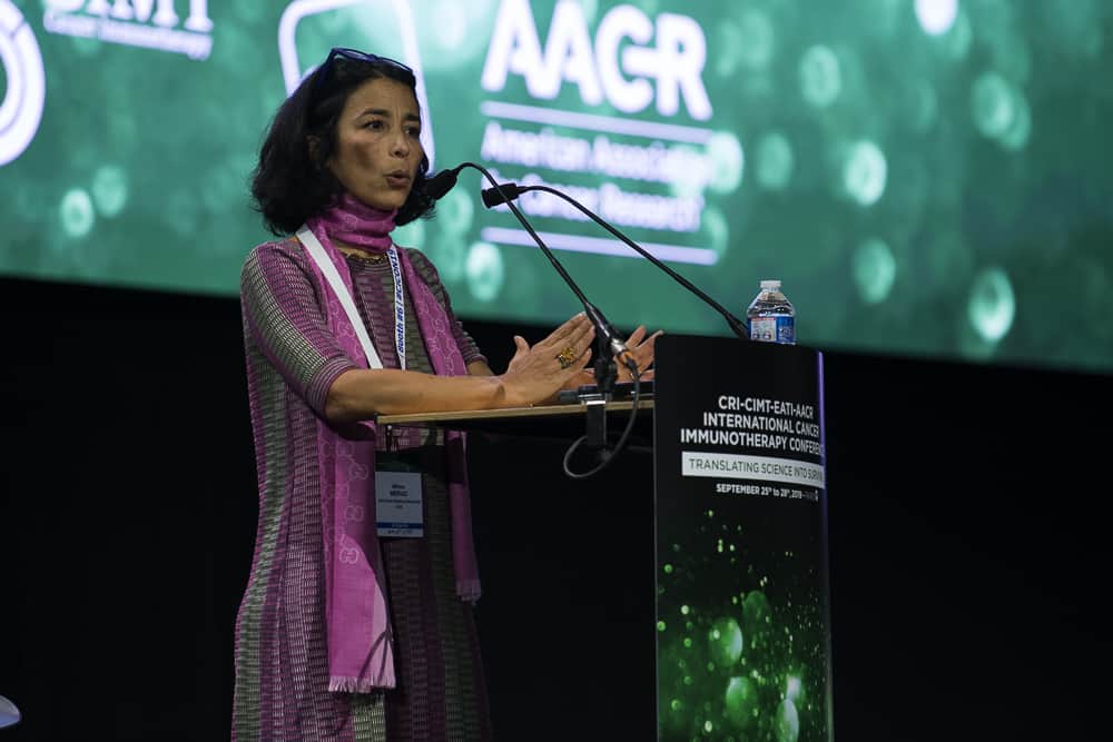
Miriam Merad, MD, PhD, discusses macrophages
As a result, we have little knowledge of the primary macrophage populations within tissues. Using a fate-mapping approach developed in her lab, she found evidence that seemed to support the existence of these two distinct macrophage lineages. Furthermore, bone marrow-derived macrophages were unable to differentiate into tissue-resident macrophages even in inflamed tissue. In the context of cancer, these tissue-resident macrophages were the first to associate with tumors and promoted tumor invasiveness and early cancer cell metastasis (spreading) during tumor development. When these tissue-resident macrophages were depleted, it reduced the intratumoral accumulation and functionality of immunosuppressive regulatory T cells, leading to better outcomes in preclinical models. Meanwhile, the presence of bone marrow-derived macrophages correlated strongly with dysfunctional T cells in tumors. One potential target found on these bone marrow-derived macrophages, but not the tissue resident type, is TREM2. The deletion of the gene encoding TREM2 led to decreased growth in lung cancer tumors and enhanced anti-tumor immunity.
The final speaker of the third day of CICON19 was Barbara B. Maier, PhD, a postdoctoral fellow in Merad’s lab, who discussed another type of myeloid cell, the dendritic cells that direct overall immune responses. One form of dendritic cells, referred to as DC-1, specialize in cross-presenting antigens to “killer” T cells and promoting their activation. As a result, these dendritic cells are important for controlling tumor growth as well as responses to immunotherapy.
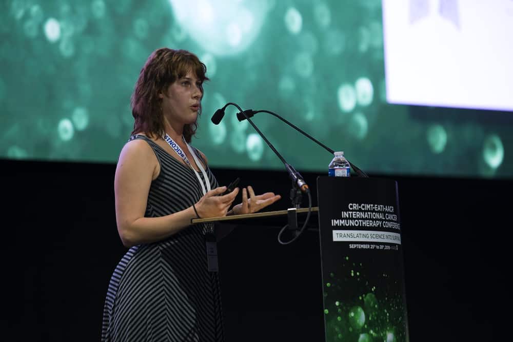
Barbara B. Maier, PhD, discusses dendritic cells
Maier also spoke about a distinct subset of DCs enriched in regulatory molecules, which she referred to as mregDCs, which are induced upon tumor antigen uptake by DCs in cancerous tissue. Additionally, these mregDCs were characterized by the production of the immune-stimulating IL-12, enhanced migratory ability (associated with increased expression of CCR7), and were also enriched in tumor-draining lymph nodes. While mregDC production of the immune-stimulating IL-12 could be dampened through IL-4 signaling, blockade of IL-4 was able to increase T cell activation and significantly reduce tumor growth.
Check our blog tomorrow for our recap of the final day of CICON19
Our complete CICON19 coverage:
- CICON19 Day 1 Update: Cancer Prevention and Combination Immunotherapy Strategies
- CICON19 Day 2 Update: T Cell Exhaustion, the Tumor Microenvironment, and Novel Cellular Immunotherapies
- CICON19 Day 3 Update: Vaccines and Tumor Antigens, Technology and Informatics, and the Tumor Microenvironment
- CICON19 Day 4 Update: Metabolic Crosstalk and the Microbiome
All photos by Arthur N. Brodsky, PhD, for the Cancer Research Institute