Immunotherapy has transformed treatment of more than a dozen types of advanced cancers, and it’s helped many patients whose disease was unlikely to respond to any other treatment. While immunotherapy doesn’t yet work for everyone, a number of promising new approaches are being developed and studied for their ability to engage the immune system and help it to eliminate cancer.
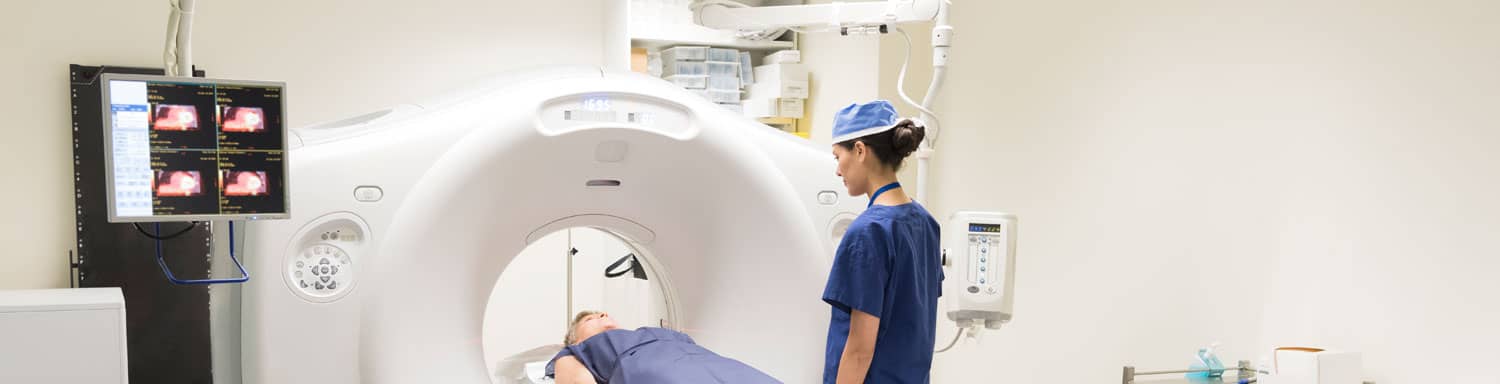
 Predicting which patients will benefit from immunotherapy and which may not is crucial to informing treatment decisions. To discuss this, we invited Kim A. Margolin, MD, an oncologist at the City of Hope National Medical Center in Los Angeles, who shared how advances in positron emission tomography (PET) scan imaging technologies may provide a more accurate picture of whether or not immunotherapy is working or is likely to work in individual cancer patients.
Dr. Margolin is a clinical professor in the department of medical oncology and therapeutics research at the City of Hope, where she serves as a principal investigator on an immunotherapy trial that is testing a new immune-based imaging technology. In addition to being triple board-certified in internal medicine with subspecialties in medical oncology and hematology, she is a fellow of the American College of Physicians and has been the chair of the Cancer Education Committee and a member of the Nominating Committee of the American Society of Clinical Oncology (ASCO). She also previously served as a member of the Oncologic Drug Advisory Committee (ODAC) of the U.S. Food and Drug Administration.
Below are some of the topics Dr. Margolin raised in our webinar, “Imaging in Immunotherapy: Using PET Scans to Guide Cancer Treatment.”
PREDICTING IMMUNOTHERAPY RESPONSES
A key question doctors face when considering treating a cancer patient with immunotherapy is whether or not a person is likely to respond to the treatment. Biopsies, or tissue samples, taken from a patient’s tumor tissue may be analyzed to see if there are biomarkers that may indicate whether a patient is more likely to benefit from the treatment.
In the case of checkpoint immunotherapy, the most broadly effective immunotherapy approach to date, there is one biomarker that seems to be especially indicative of potential responses: the presence of “killer” T cells within tumors. Patients whose tumors have been infiltrated by these immune cells are much more likely to respond to checkpoint immunotherapy. Surgical biopsies, however, are by definition an invasive procedure that comes with some safety risks. Fortunately, incorporating immune-based technologies into PET scans, which are already used to get basic information such as tumor size and location, may enable doctors to see immunological activity inside tumors in a safer, quicker, and less invasive way.
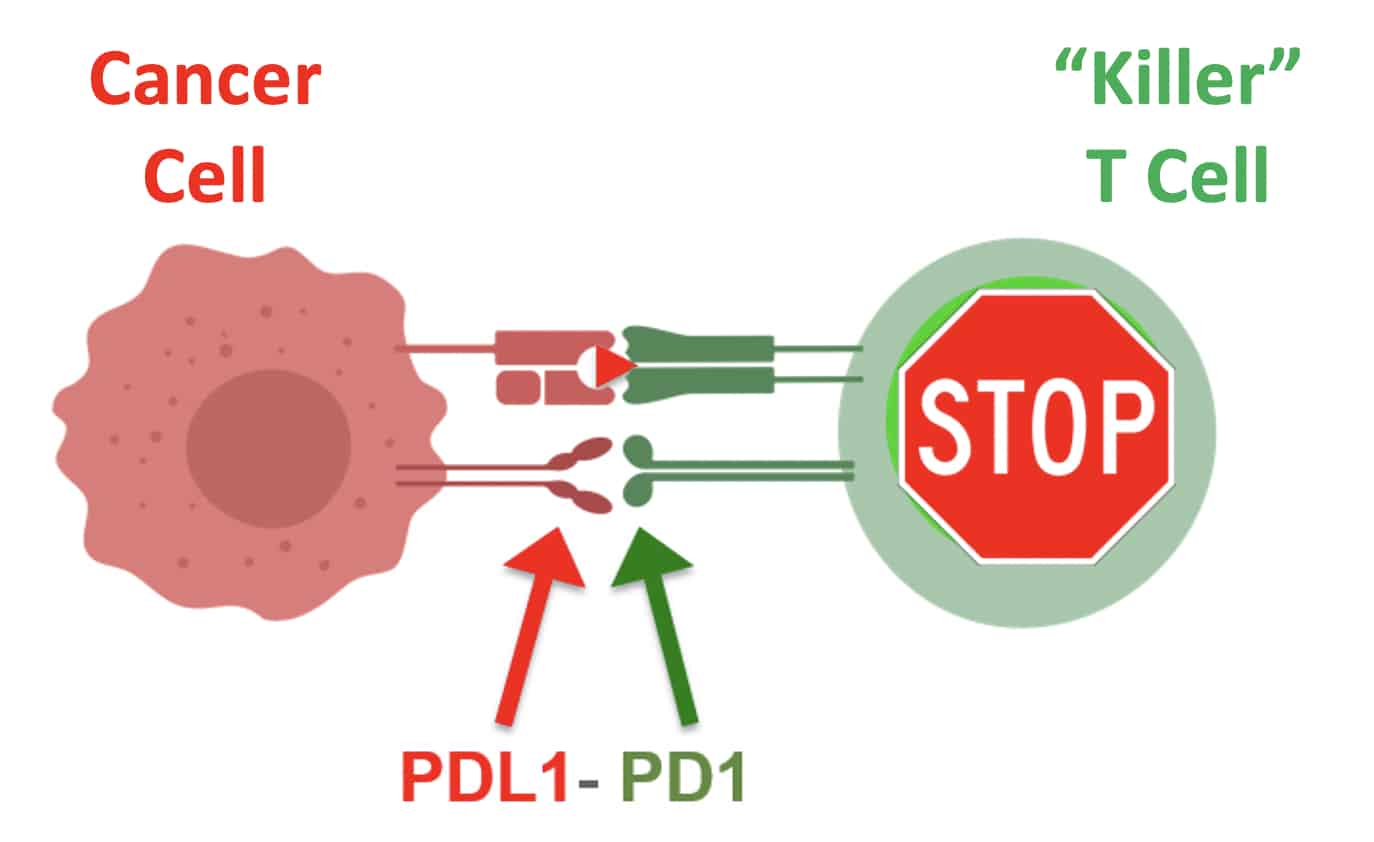
An immune checkpoint illustrated.
USING PET SCANS TO PREDICT IMMUNOTHERAPY RESPONSES
Dr. Margolin discussed a phase 2 clinical trial that she’s currently leading in which PET scans are being used to detect killer T cells within the tumors of patients with metastatic cancer who are being treated with checkpoint immunotherapy and chemotherapy.
One of the most important components of killer T cells is the CD8 receptor that they express on their surface. This receptor enables T cells to interact with and kill cancer cells or any other damaged or diseased cell in our body.
In this trial, as Margolin highlighted, her team is using a specialized molecule called an engineered antibody fragment. Antibodies are a key component of the immune system that flag danger or directly neutralize it. The engineered antibody fragment Dr. Margolin is testing in the clinical trial is designed to target the CD8 receptor on killer T cells, but contains a radiolabeled particle that allows her to detect the presence of the killer T cells. This low-level radioactive signal can be measured by the PET scan, and in turn, allows a patient’s T cells—if there are enough of them—to be visualized within their tumors via a non-invasive PET scan.
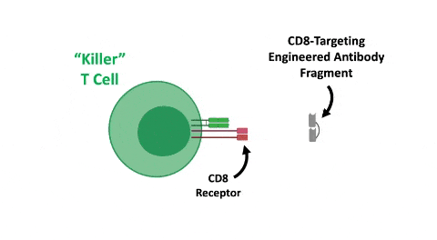
CD8-targeting engineered antibody fragment attaches to killer t cell and creates a signal.
While the presence of T cells within a tumor is promising, it in no way guarantees a patient will respond to checkpoint immunotherapy. As Margolin discussed in the webinar, their presence in the tumor doesn’t necessarily mean that they are all cancer-targeting T cells designed to recognize tumor markers. At the moment, it’s also unclear whether there is a minimum amount of cancer-targeting T cells required to effect a successful response to immunotherapy. Much more detail about this connection remains to be characterized.
In addition to the detection of T cells within tumors, PET scans have a variety of other applications in immunotherapy. For instance, engineered antibody fragments have been developed to target (and enable the measurement of) the PD-L1 protein, another useful biomarker in the context of PD-1/PD-L1 immunotherapy. Cancer-specific markers can also be targeted and visualized to gain a better understand of the tumor’s behavior. One example is the HER2 protein that is abnormally overexpressed in a variety of cancers. Knowing whether a cancer expresses high levels of HER2 may help guide treatment-related decisions with respect to a variety of immunotherapies that target HER2.
THE FUTURE OF IMAGING IN IMMUNOTHERAPY
Margolin spoke about how immunoPET technology may impact cancer treatment in the future. This technology provides a safe and non-invasive way to look inside patients’ tumors and determine whether their immune systems are responding. Of course, the most immediate practical application is to guide decisions regarding current treatments, either in recently diagnosed or treatment-resistant patients. Dr. Margolin emphasized that these technologies won’t necessarily see widespread adoption—especially at the community level—anytime soon. More testing must still be done to validate their utility. Moving forward, immune-based PET imaging could also enable doctors to monitor patients over time in response to various treatments. This would allow doctors to know if a treatment is working, or if it might be time to consider potential next options for the patient.
In the longer term, enabling easier study of immune response to tumors over time in both people and animals may help improve our understanding of a variety of immune-related processes within the context of cancer. This includes in-depth characterization of a variety of immune cell populations, including B cells, regulatory T cells, macrophages, natural killer cells, and dendritic cells. These insights could ultimately reveal new mechanisms that are crucial for successful anti-cancer immune responses and provide a rationale for the development of next-generation immunotherapies that target these cells and pathways.
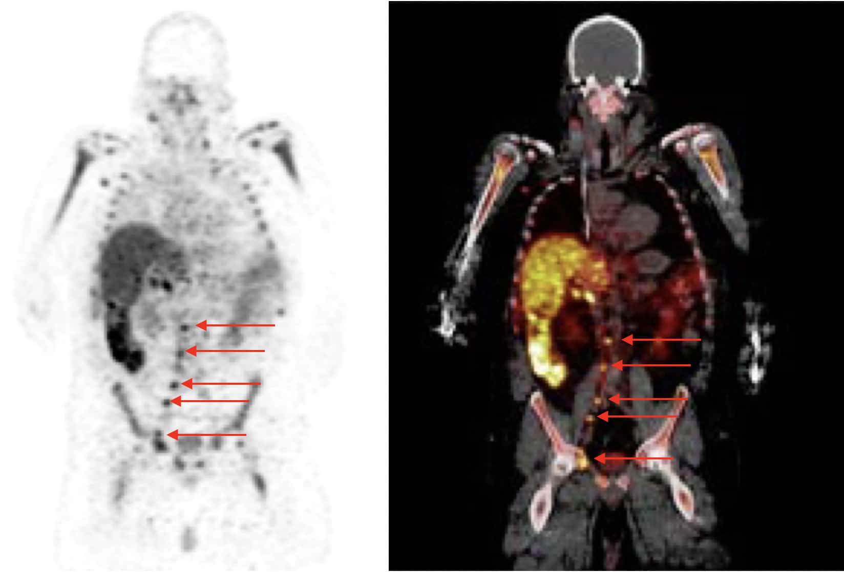
Patient CD8 PET scan
This webinar was a special edition of CRI’s “Cancer Immunotherapy and You” webinar series produced by the Cancer Research Institute and was hosted by our director of marketing and communications, Brian Brewer. Sponsorship for this webinar was generously provided by ImaginAb. Sponsorship does not influence editorial decisions or content. Browse our Cancer Immunotherapy and You Webinar Series playlist on YouTube or visit the Webinars page on our website to see other webinars in this series.